Revolutionizing Pediatric Cardiology: How Children’s Hospital of Philadelphia and Open-Source AI Are Transforming Congenital Heart Surgery Planning

Congenital heart disease remains one of the most persistent and delicate challenges in modern pediatrics. Affecting approximately 1% of all live births, congenital heart defects are as varied as the children who are born with them. No two pediatric hearts are identical, presenting a staggering anatomical puzzle for pediatric cardiologists and cardiovascular surgeons. Historically, clinicians treating these fragile patients have relied on off-the-shelf surgical devices and standard-issue components designed for broad anatomical averages, forcing them to adapt generic tools to entirely unique, miniature biological systems.
This paradigm is undergoing a fundamental transformation at the Children’s Hospital of Philadelphia (CHOP), where a dedicated team of researchers and clinicians is leveraging cutting-edge open-source artificial intelligence and advanced physics simulation tools. By generating hyper-precise, patient-specific 3D heart models in mere seconds, CHOP is pioneering a new era of personalized medicine that aims to significantly enhance surgical precision, reduce operating times, and dramatically improve outcomes for children born with complex heart defects.
The Core Innovation: Speed Meets Anatomical Precision
At the heart of CHOP’s cardiac modeling service is MONAI, an open-source medical imaging AI framework co-founded by NVIDIA. By processing routine diagnostic imagery—such as computed tomography (CT) scans, magnetic resonance imaging (MRI), and specialized 3D echocardiography—the technology translates complex two-dimensional datasets into high-fidelity, three-dimensional anatomical models.
Previously, generating a single, reliable heart model required a skilled researcher to spend up to four hours manually segmenting tissues and mapping structures at a dedicated workstation. While effective for retrospective research, that time-intensive workflow made routine clinical adoption impractical for fast-moving surgical teams. Through the integration of MONAI Label and NVIDIA’s Auto3DSeg implementation, Dr. Matthew Jolley and his research team at CHOP have trained advanced neural networks on historical pairs of clinical images and validated models. Today, the system matches the precision of human specialists, producing accurate diagnostic models in a matter of seconds.
This newfound computational speed has allowed cardiac modeling to transition smoothly from an experimental research application into an active, everyday component of clinical care. For complex pathologies such as ventricular septal defects—commonly referred to as holes between the lower chambers of the heart—modeling is increasingly becoming a standard preoperative requirement. Surgeons can now virtually inspect a patient’s exact internal geometry, evaluate structural anomalies that traditional imaging fails to clearly resolve, and simulate interventions long before the patient ever enters the operating room or catheterization laboratory.
A Decadelong Journey from Research Bench to Clinical Bedside
The integration of advanced computational modeling into pediatric cardiology is not an overnight phenomenon, but rather the culmination of a deliberate, decadelong evolution. When Dr. Jolley joined CHOP in 2015, three-dimensional echocardiography was emerging as a promising clinical tool. While robust software packages existed for modeling adult cardiac valves, virtually no commercial or academic tools were tailored to the complex, miniature anatomies characteristic of congenital heart patients.
Recognizing this gap, Jolley’s laboratory collaborated closely with the broader open-source software community to develop SlicerHeart, an specialized extension of the widely used 3D Slicer platform designed for visualizing, segmenting, and analyzing three-dimensional medical imagery. Over subsequent years, the team engineered specialized processing pipelines aimed at reconstructing pediatric hearts and valves from multimodal imaging sources.
The introduction of machine learning served as the primary catalyst that bridged the gap between academic exploration and clinical utility. By automating the tedious segmentation process through supervised training on historical patient datasets, the technology eliminated the human bottleneck. As Jolley notes, machine learning has effectively integrated into the lab’s foundational workflow; once a modest batch of ten to twenty image-model pairs is established, automated training routines rapidly deploy new segmentations into clinical pipelines.
Expanding Horizons: Real-Time Biomechanical Simulation with Newton and NVIDIA Warp
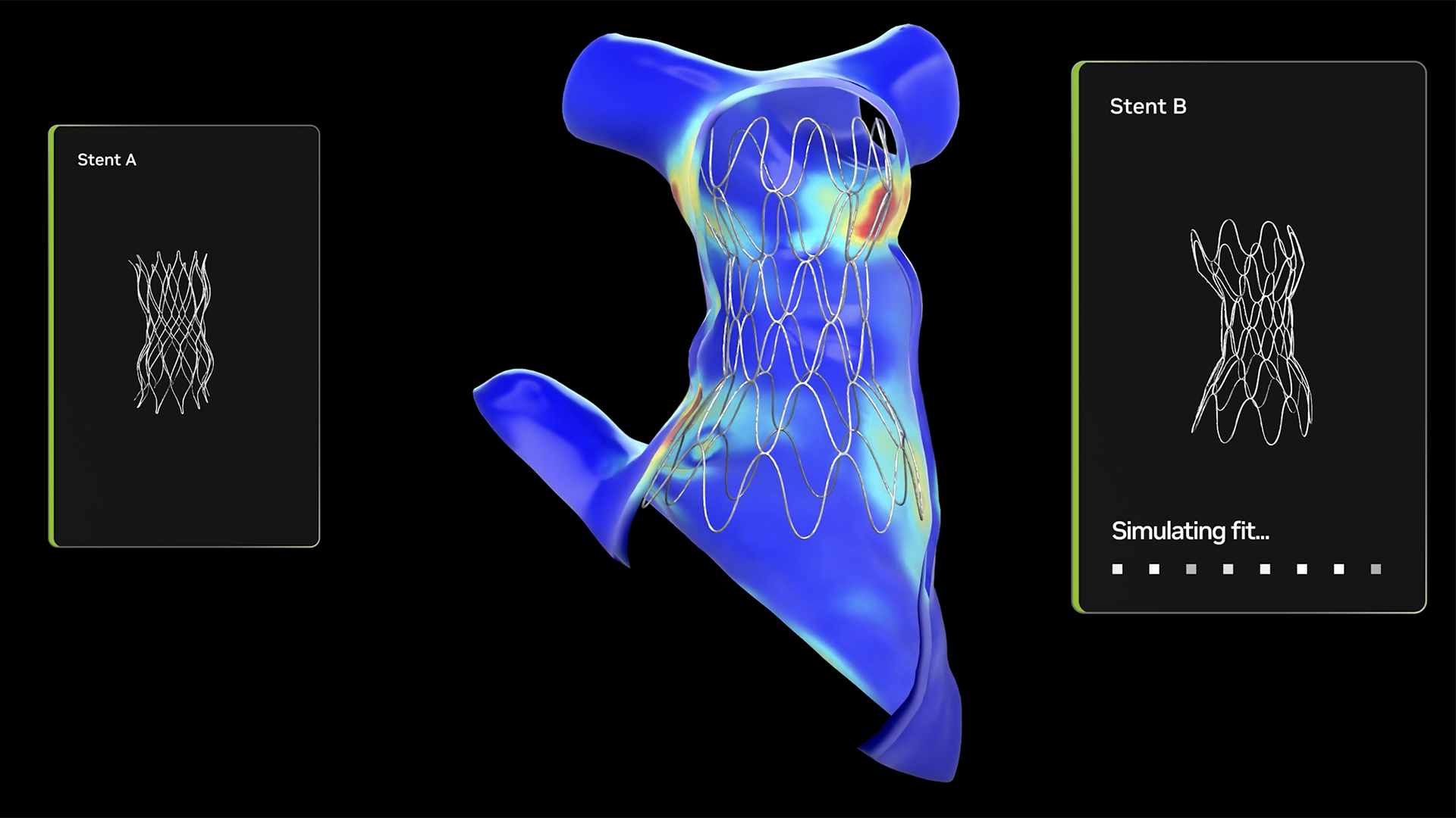
While visualizing a patient’s specific anatomy provides critical diagnostic clarity, static 3D models represent only the first phase of surgical planning. The next frontier in pediatric cardiology involves predictive biomechanics—determining precisely how living cardiac tissue will respond when a foreign device, such as an artificial valve or closure plug, is deployed inside it.
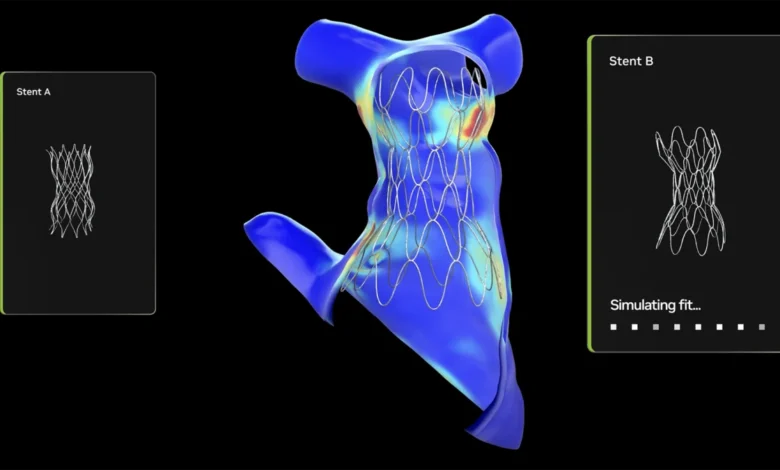
To achieve this predictive capability, CHOP has incorporated Newton, an open-source physics engine built upon the NVIDIA Warp Python framework, which executes complex physics simulations directly on graphics processing units (GPUs). In collaboration with NVIDIA and open-source contributors, CHOP is developing specialized biomechanical simulation frameworks optimized for Warp.
Historically, simulating the mechanical interaction between a cardiac device and dynamic heart tissue required hours of computational processing, often demanding overnight runs for multiple structural configurations. Leveraging GPU acceleration, the newly integrated workflows reduce simulation times to near-real-time thresholds. Clinicians can now evaluate how alternative artificial valves or occlusion devices will fit within a specific child’s anatomy, analyzing wall stress distributions and hemodynamic responses to inform same-day clinical decisions.
Looking further ahead, CHOP is actively developing integrated couplers utilizing SlicerHeart and NVIDIA Omniverse digital twins powered by OpenUSD (Open Universal Scene Description). By enabling seamless 3D data interoperability, OpenUSD allows diverse solvers and patient datasets to converge within immersive virtual reality environments. Enhanced by embedded vision-language models (VLMs), these environments will soon allow medical teams to intuitively query, visualize, and interact with complex cardiac simulations through natural language and gesture control before performing actual procedures.
Overcoming Economic Barriers Through Collaborative Open Source
The development and deployment of these advanced computational tools address a harsh economic reality inherent in pediatric healthcare. With approximately 2.4 million individuals living with congenital heart disease across the United States, the patient population is comparatively rare and highly heterogeneous. Traditional medical device manufacturers have historically found it economically unviable to invest heavily in specialized software and hardware infrastructure tailored to such niche demographic cohorts.
Open-source frameworks provide an effective counter-weight to these traditional market economics. Because foundational platforms like SlicerHeart, MONAI, Newton, and 3D Slicer are freely accessible and endlessly expandable, academic institutions and research hospitals can pool their intellectual resources without encountering proprietary commercial barriers.
A growing national consortium of leading pediatric institutions—including Boston Children’s Hospital, Stanford University, and CHOP—is actively establishing collaborative networks to share modeling infrastructure and software extensions. At Boston Children’s Hospital, cardiac modeling currently supports more than half of all surgical interventions, translating to roughly 500 cases annually. Meanwhile, CHOP anticipates scaling its internal volume to approximately 200 modeled cases this year alone, with broader institutional plans to export these AI-driven workflows into additional clinical disciplines through the IDEA Lab, housed within the Morgan Center for Research and Innovation.
Broader Implications for Specialized Medicine
The partnership between clinical pediatric research and industrial-scale open-source AI infrastructure establishes a replicable blueprint for other underserved medical specialties. By harnessing platforms maintained by robust developer ecosystems, individual children’s hospitals can command computational capabilities that would traditionally require massive, enterprise-level engineering teams to build and maintain independently.
As these tools mature and integrate deeper into standard clinical pathways, the implications for patient safety and procedural efficacy are profound. By shifting from generalized, population-based assumptions to individualized, physics-simulated precision medicine, pediatric cardiologists are steadily redefining the boundaries of what is possible. For the children and families navigating the complexities of congenital heart disease, open-source innovation is turning uncertainty into actionable, life-saving clarity.